*手术步骤
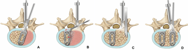
Figure 1.
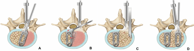
双通道内镜经椎间孔腰椎椎体间融合术(Biportal Endoscopic Transforaminal Lumbar Interbody Fusion, BETLIF)中双融合器植入的手术步骤。
(A) 将第一个融合器以斜向方式插入,并深入至对侧椎间隙。
(B) 在内镜视野下重新调整融合器的位置。
(C) 可使用漏斗(funnel)将骨移植物打压植入椎间隙。
(D) 在保护同侧神经根的同时,将第二个融合器以垂直方向植入。
*方法
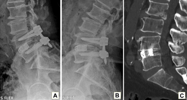
Figure 2.
术后6个月及1年,通过动态X线平片和计算机断层扫描(Computed Tomography, CT)进行融合评估,以判断是否达到成功融合。
(A, B) 术后6个月屈曲—伸展位动态X线片。
(C) 术后6个月CT扫描。
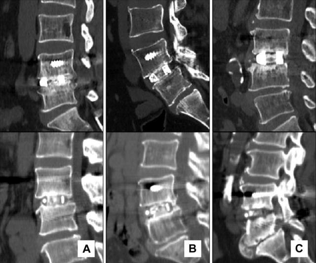
Figure 3.
采用 Bridwell grading system(Bridwell分级系统) 评价融合等级。
下排为 PEEK融合器,上排为 3D打印钛融合器(3D-printed titanium cage)。
融合等级分别为:
(A) Grade I
(B) Grade II
(C) Grade III
融合情况按照Grade I至Grade IV进行分级:
* Grade I: 已实现融合,可见骨重塑及骨小梁形成。
* Grade II: 骨移植物保持完整,尚未完全重塑和整合,但未见透亮区(lucency)。
* Grade III: 骨移植物保持完整,但移植物上、下缘可能存在透亮区。
* Grade IV: 未实现融合,并伴有骨移植物塌陷或吸收。
根据影像学结果,在Bridwell grading system中,Grade I和Grade II被定义为solid fusion(坚固融合)。
本研究中未出现任何Grade IV病例。
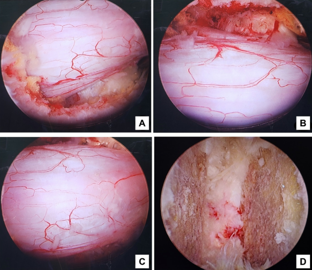
Figure 4.
双通道内镜经椎间孔腰椎椎体间融合术(BETLIF)的术中内镜图像。
(A) 右侧同侧下行神经根(ipsilateral traversing nerve root)充分减压后的状态。
(B) 对侧下行神经根(contralateral traversing nerve root)充分减压后的状态。
(C) 中央椎管(central canal)充分减压后的状态。
(D) 内镜下终板处理(endplate preparation)图像。使用剥离器(dissector)将软骨终板(cartilaginous endplate)与骨性终板(osseous endplate)分离。
*结果
术后1年随访时,3D打印钛融合器组的融合等级分别为:
Grade I:75.0%(36个节段)
Grade II:20.8%(10个节段)
Grade III:4.2%(2个节段)
PEEK融合器组的融合等级分别为:
Grade I:53.2%(25个节段)
Grade II:40.4%(19个节段)
Grade III:6.4%(3个节段)
两种融合器的总体融合率(Grade I和II)相近,分别为 95.8%和93.6%(P = 0.629),差异无统计学意义。
然而,与PEEK融合器相比,3D打印钛融合器组的 Grade I融合比例显著更高(75.0% vs 53.2%,P = 0.027)。
两种融合器在**融合器沉降(subsidence)及并发症(complications)**方面均未观察到显著差异。
多变量分析(multivariate analysis)显示,年龄是BETLIF术后融合器沉降唯一具有显著相关性的变量。
*结论
双3D打印钛融合器和双PEEK融合器均表现出较高的融合率,在总体融合成功率方面两者无显著差异。
然而,与PEEK融合器相比,双3D打印钛融合器具有更好的早期融合等级,同时其融合器沉降情况与PEEK融合器相当。
尽管仍需长期随访以进一步确定其疗效,但本研究结果提示,在BETLIF中,3D打印钛融合器在早期融合质量方面可能具有优势。
未来仍需进一步研究,以优化融合器排列方式(cage arrangement)、融合器设计(cage design)及手术技术(surgical techniques),从而进一步改善临床疗效。
*临床相关性
推荐在BETLIF中使用双3D打印钛融合器(double 3D-printed titanium cages)。
DH LEE et al. Use of Double Cages for Biportal Endoscopic Transforaminal Lumbar Interbody Fusion: A Comparison of 3-Dimensional-Printed Titanium and Polyetheretherketone Cages.
Int J Spine Surg published online 29 August 2025
-----
英文原版
Use of Double Cages for Biportal Endoscopic Transforaminal Lumbar Interbody Fusion:A comparison of 3D Titanium and PEEK Cages
SURGICAL STEP
Figure 1.
Surgical steps of 2-­cage insertion during biportal endoscopic transforaminal lumbar interbody fusion. (A) The first cage is obliquely inserted deeply into the contralateral disc space. (B) The cage is repositioned under endoscopic view. (C) A funnel may be used to impact bone transplants into the disc space. (D) The second cage is inserted vertically while protecting the nerve roots in the ipsilateral direction.
Methods
Figure 2.
Fusion evaluation was used to assess a successful fusion with dynamic plain radiography and computed tomography (CT) at 6 mo and 1 y postoperatively. (A and B) Flexion-­extension plain x-­ray images at 6 mo postoperatively. (C) CT scan at 6 mo postoperatively.
Figure 3.
The Bridwell grading system is used to evaluate fusion grade. The bottom row is a PEEK cage, while the top row is a 3-­dimensional–printed titanium cage. The grades are (A) grade I, (B) grade II, and (C) grade III. Fusion is ranked from I to IV: I represents fusion with remodeling and trabeculae; II represents graft intactness, not entirely remodeled and integrated, but without lucency; III represents graft intactness, with possible lucency at the top and bottom of the graft; and IV represents fusion absence with graft collapse or resorption. Solid fusion is classified as grade I and II in the Bridwell grading system based on radiological results.
The present study did not have any grade IV data.
Figure 4.
Intraoperative endoscopic images of biportal endoscopic transforaminal lumbar interbody fusion. Fully decompressive status of the right ipsilateral traversing nerve root (A), contralateral traversing nerve root (B), central canal (C), and endoscopic endplate preparation image (D). The cartilaginous endplate is separated from the osseous endplate using a dissector.
Results
At 1-­year follow-­up, fusion grades were I (75.0%, 36 levels), II (20.8%, 10 levels), and III (4.2%, 2 levels) for 3D-­printed titanium and I (53.2%, 25 levels), II (40.4%, 19 levels), and III (6.4%, 3 levels) for PEEK. The overall fusion rate (grades I and II) was similar for both cages (95.8% vs 93.6%, P = 0.629), but grade I was more prevalent with 3D-p ­ rinted titanium than with PEEK (75.0% vs 53.2%, P = 0.027). No significant differences were observed in subsidence or complications between the 2 cages. Multivariate analysis revealed age as the only variable significantly associated with subsidence in BETLIF.
Conclusions
Both double 3D-PRINTED TITANIUM AND PEEK CAGES demonstrated high fusion rates with no significant differences in overall success. However, double 3D-­printed titanium cages showed better early fusion grades and comparable subsidence to that of PEEK cages. Although long-­term follow-­up is necessary to ascertain efficacy, these findings suggest that 3D-­printed titanium cages offer advantages in early fusion quality in BETLIF. Further research is needed to optimize cage arrangement, cage design, and surgical techniques to improve outcomes.
Clinical Relevance: The use of double 3D-­printed titanium cages is recommended in BETLIF.